Esthetic treatment of severely damaged teeth (i.e. over 50% loss of tooth structure) for crown and bridgework in the anterior region requires tooth-coloured materials and a post to improve the retention of the core build up.
Glass fibre-reinforced or ceramic post systems are the first step in creating adequate retention for the subsequent core build up and all-ceramic crown. The question regarding which type of post should be given preference - ceramic or fibre reinforced, is a controversial subject in the literature.Glass fibre-reinforced posts have the distinct advantage of providing better distribution of the stress forces within the residual tooth structure due to their dentine-like modulus of elasticity. Adhesive cementation of the post to the endodontically treated dentine should increase the retention of the core build up and improve the distribution of stress forces during loading; thus preventing the risk of root fracture (Freedman et al. 2008).
From a pure mathematical standpoint, if fewer dissimilar materials are used for adhesive cementation, fewer varying interfaces will exist. Consequently, there will be fewer weak points created when restoring the tooth root with a post. The goal of the clinical case documented in this report was to cover all of the steps in the treatment using a single adhesive system. The post was therefore cemented using ParaCore and ParaBond – a chemically cured adhesive. On the other hand, the core build up was cemented using ParaCore only – the contact surfaces can also be conditioned with adhesive when preparing the root canal. The crown was also cemented using ParaCore and ParaBond. The bond strength and flexural strength (composite is ideal for this) between the tooth, ParaCore, post and crown material should attain optimal values. Laboratory tests by Millar et al. 2008 demonstrated the validity of the theory behind the ParaCore System.
Dye penetration tests produced evidence of an excellent seal and an optimal cement margin. This indicated more efficient bonding than other systems tested in this study.
The author surmised that the chemical adhesive was the key to the good test results.
This in turn points to the excellent properties of ParaCore in the required areas of application, such as post cementation, core build-up and contouring, and finally, cementation of the restoration.
The use of one material for all three indications produces the desired “monoblock bond interface”. The clinical procedure of canal preparation, fitting of the post and cementation, etc. does not differ from other existing post systems. Treatment using only one material for all three indications enables a costeffective as well as time-saving procedure. Maximum stability is attained due to the resulting “monoblock bond interface”.
The procedure is demonstrated step by step in the following clinical case presented.
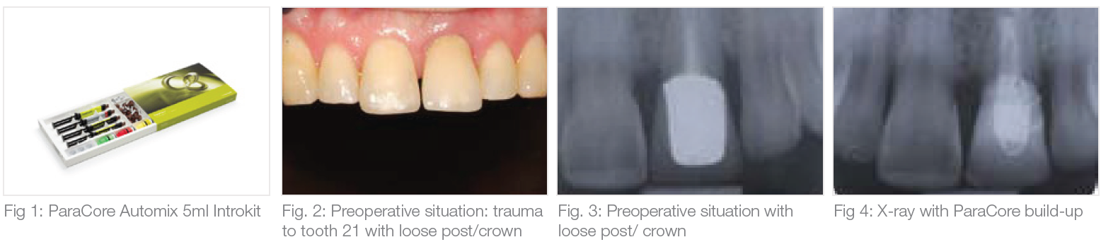
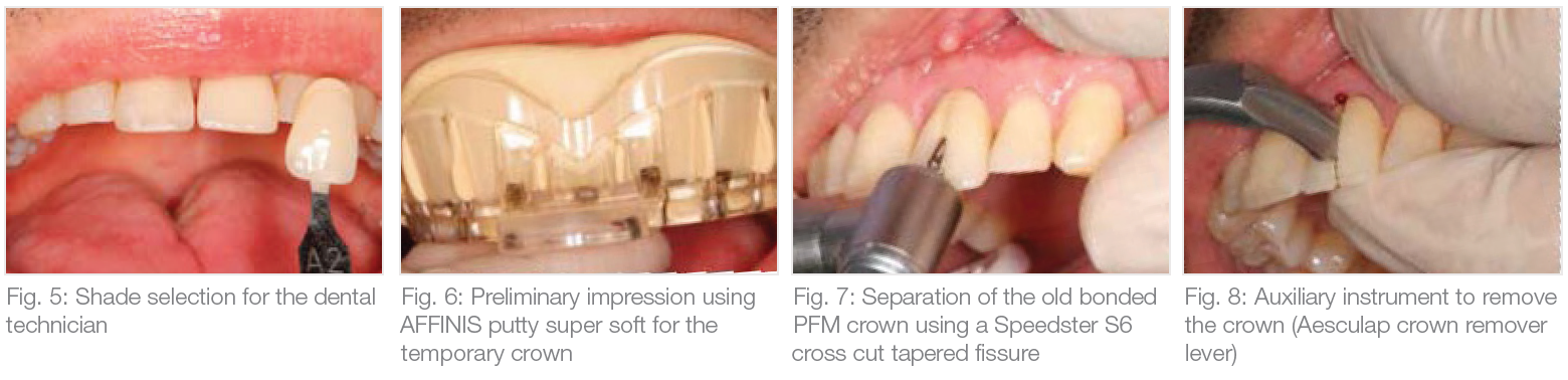
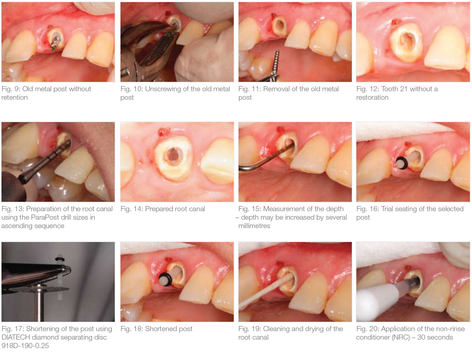
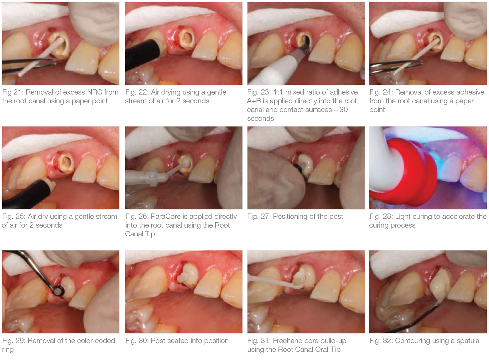
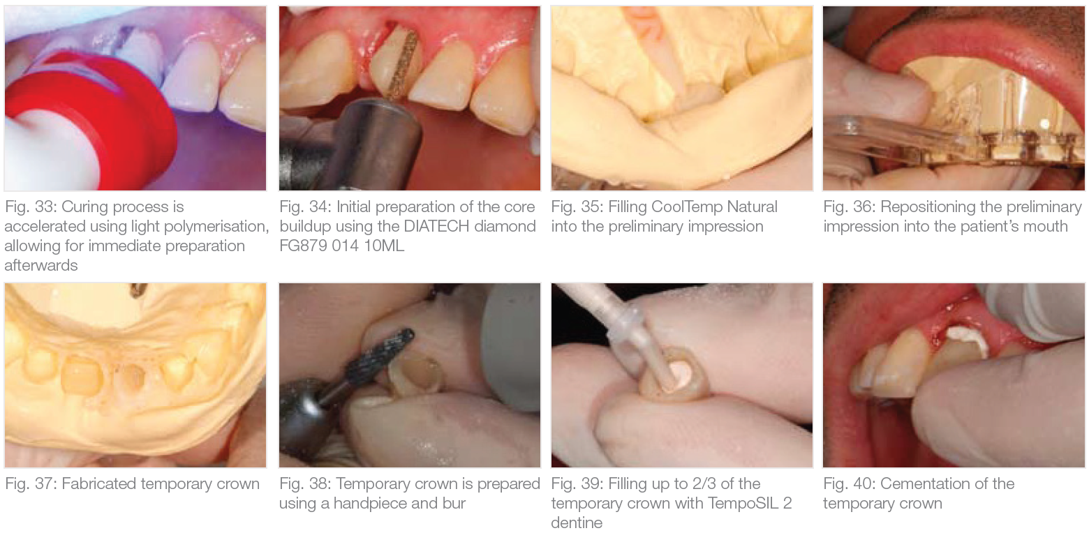
Due to a trauma-related incident to his anterior tooth 21 during his adolescent years, the patient had several prior treatment procedures performed and presented in the practice with a loose post crown (Fig. 2). There were no fracture lines visible on the x-ray with only a slight deviation of the prepared post canal inside the root canal (Fig. 3). This deviation was incorporated when the new post was fitted (Fig. 4). After the shade selection (Fig. 5) and preliminary impression (Fig. 6), the old crown with the metal framework and metal post, which was loose inside the root canal, were removed (Fig. 7–12). The root canal was then prepared using ParaPost Drill sizes in ascending sequence (Fig. 13+14). The prepared depth was measured using a periodontal probe (Fig. 15). The length of the Fiber Lux selected in this case had to be shortened (Fig. 16–18). After the preparation was completed, the root canal was cleaned (Fig. 19), and ParaBond adhesive was applied into the root canal and onto other contact surfaces (Fig. 20–25). Simultaneous conditioning of the contact surfaces eliminates re-application of the adhesive during the core build-up. Use of a rubber dam, which is normally routine practice, was not required because of the clinical situation. The Root Canal Tip of ParaCore can be used for direct application into the root canal (Fig. 26). Once the post was positioned into the root canal (Fig. 27), it was secured into position using a curing lamp (Fig. 28). The core build up prepared using the freehand technique and completed using additional polymerization steps (Fig. 29–34).
Clinical tip: material stored in the refrigerator extends the working time, which can be particularly practical in summer. Following initial preparation, a temporary crown was fabricated using CoolTemp Natural (Fig. 35–38) and cemented using TempoSIL 2 (Fig. 39–41).
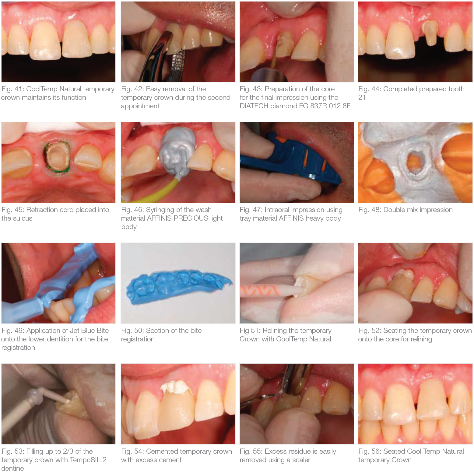
The temporary crown was easily removed during the second appointment (Fig. 42) with the preparation finalised afterwards (Fig. 43+44). A one-step impression was taken using AFFINIS PRECIOUS light for reproduction of very crisp detail, and AFFINIS heavy body tray material.
Both are addition-curing silicones (Fig. 45–48). After preparing the bite registration (Fig. 49+50), the direct temporary crown was relined (Fig. 51+52) and recemented using TempoSIL 2 (Fig. 53–56).
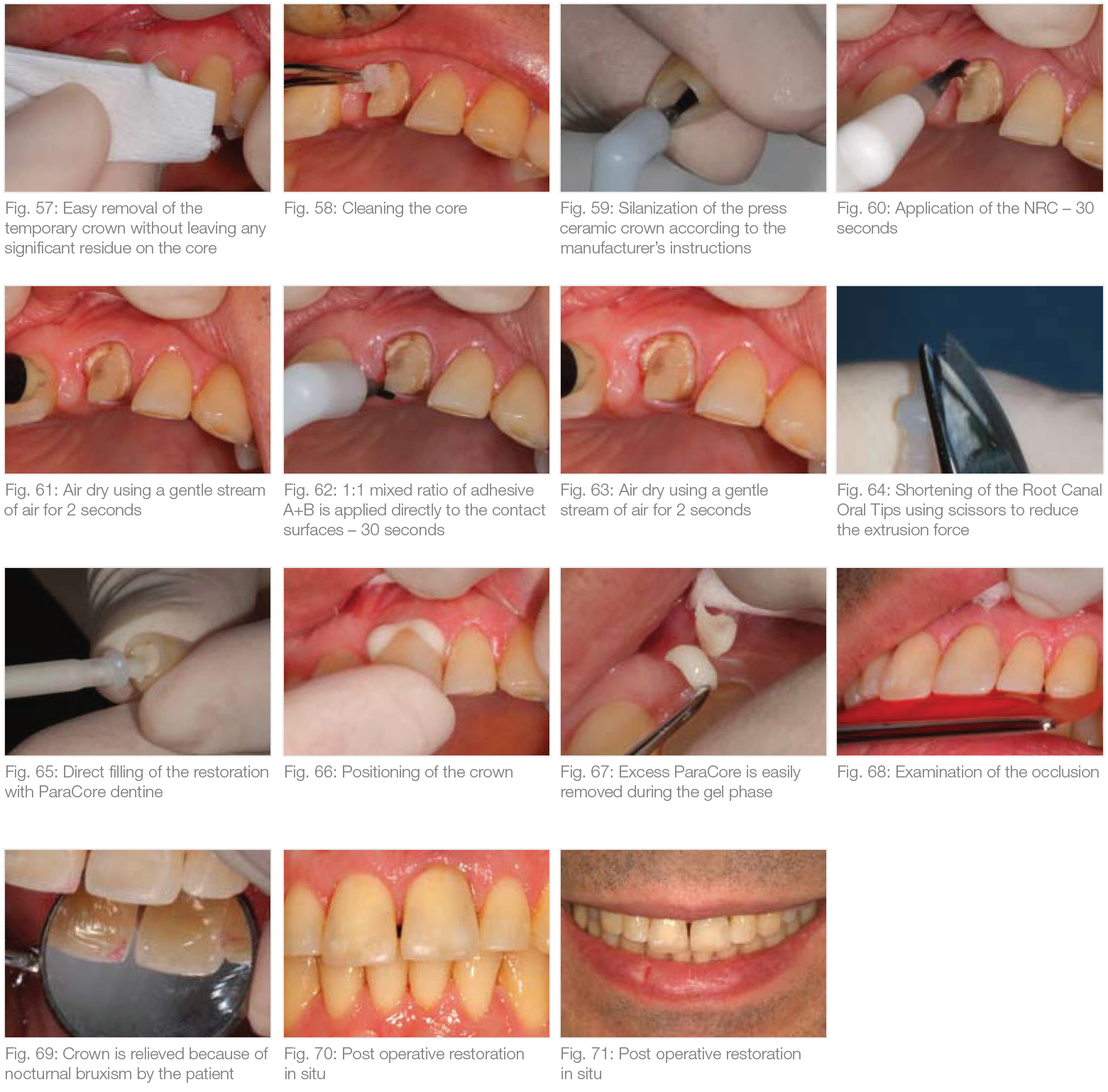
After completing all of the necessary preparations (Fig. 57–59), the all-ceramic crown fabricated by the dental technician was adhesively cemented using the ParaBond/ParaCore System (Fig. 60–66). Two shades are available for cementation, white and dentine. The advantages of this product become particularly evident with all-ceramic anterior crowns ensuring for perfect integration of the shade to the dentition.
After the fitting of the crown, excess residue was removed during the gel-like curing phase of the material (Fig. 67).
Excess residue can also be easily removed using a scaler, once the cement is completely cured. After checking the occlusion, treatment was complete and the patient left the practice with a new restoration (Fig. 68–71).
The instructions for use (step-by-step cards) supplied with the ParaCore System are simple and user friendly to ensure that these procedures are quickly integrated into your daily practice routine.
With ParaCore, one material is now available to accommodate all of the prescribed steps for cementation of an anterior crown using one material for a monoblock bond interface.


DR. CHRISTOPH G. HÜSKENS
MED. DENT. October 2009