Drs Carlos Colino, Laura Castillo, Isabel Drewling & Pedro Colino, Spain
Fig. 1: Initial extra-oral photographs.
Introduction
Clear aligners are increasingly being used by orthodontists and requested by patients for its aesthetics, better hygiene and comfort. Thanks to biomechanical advances and materials, the effectiveness of treatments has improved.1, 2 In addition, very promising results have been shown in Class II correction by distalisation of maxillary molars together with Class II elastics and very good control of the vertical dimension in cases of deep overbite or open bite.3, 4 Sequential distalisation is a good means of correcting Class II malocclusion and can be combined with posterior interproximal reduction and elastics to improve efficiency, but it involves an average of 12 aligners (three months if we change the aligners every week) only to perform the 2–4 mm distalisation of a first and second molar.5
Although sequential distalisation is highly predictable, it constitutes a treatment that involves many stages, the extent of which varies depending on the severity of the Class II malocclusion. Nowadays, patients desire not only aesthetic results but also fast results, as they tend to lose patience over the course of treatment, especially adolescents. For this reason, we have opted for a hybrid approach consisting of a first phase using the Carriere Motion Appliance (Henry Schein Orthodontics) to correct Class II malocclusion within a span of four to five months and a second phase during which aligners are used once a Class I occlusion has been established, thus reducing the number of aligners considerably and allowing for shorter treatment times. We have named this approach the minimal time orthodontics (MTO) system, as it not only reduces the frequency of appointments with the patient, but also shortens the total treatment duration.
Diagnosis
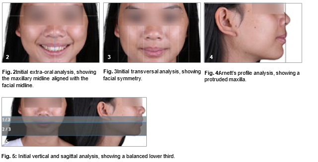
A 15-year-old mild brachyfacial female patient in Stage 5 of cervical maturation presented with a skeletal and dentoalveolar Class II, Division I malocclusion due to maxillary protrusion. The patient’s main concern was the increased overjet and dental exposure when smiling—she was being bullied. She had permanent dentition and good oral hygiene. In the extra-oral analysis (Figs. 1 & 2), we observed facial symmetry (Fig. 3), mandibular retrusion (Fig. 4) and a vertically balanced lower third (Fig. 5).
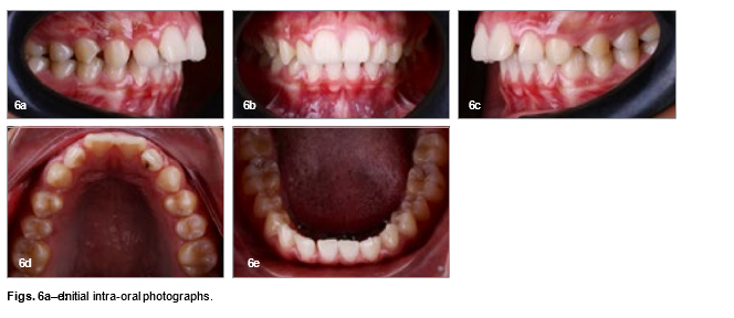
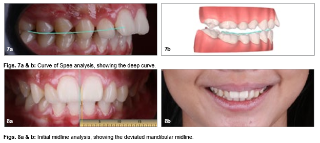
In the intra-oral analysis, we observed a bilateral Class II molar and canine relationship, along with pronounced diastemas between the premolars and canines (Fig. 6). There was a significantly increased overjet, which became very evident during smiling, and a deep lower curve of Spee (Fig. 7), and the mandibular midline deviated 0.5 mm to the left of the maxillary midline (Figs. 8–11). We use the iTero intra-oral scanner (Align Technology).
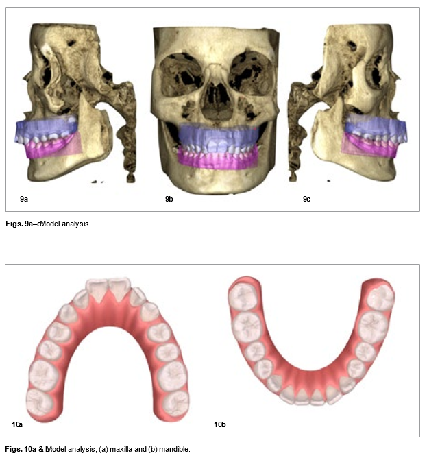
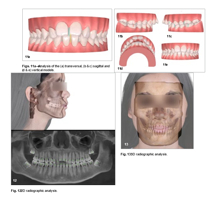
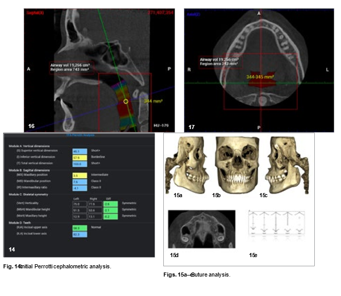
The dental panoramic tomogram revealed that the four third molars had not erupted (Fig. 12). We used the 3D scan to complete the diagnosis (Fig. 13). The Perrotti total face approach cephalometric analysis was conducted with the Planmeca Romexis software, and it found that the patient was slightly brachyfacial and skeletally symmetrical and presented with a skeletal Class II malocclusion due to mandibular retrusion (Fig. 14). She exhibited maturation Stage C of the mid- palatine suture (Fig. 15),6 no transverse maxillary concerns and a good airway volume of 19.3 cm3 (Figs. 16 & 17).
Treatment
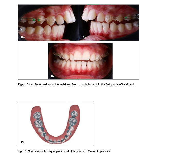
Our treatment focused on solving the issues identified in the three planes of space using the Sagittal First approach with Carriere Motion Appliances and clear aligners (Reveal, Henry Schein Orthodontics). We conducted digital planning using the Reveal software to design the mandibular aligners. These aligners are intended to provide additional negative torque on the mandibular incisors, thereby counteracting the adverse effects of the Carriere Motion Appliances due to elastic use (Fig. 18). Additionally, these aligners served as a retention method after the standard Carriere Motion Appliance protocol. We do not schedule patient appointments for placement of Carriere Motion Appliances until we have the mandi- bular aligners in our office.
In approximately 15 minutes, we completed the cementation of the Carriere Motion Appliances and provided patient education regarding the 22-hour daily elastics protocol. The protocol was 6 oz elastics during the first month and then 8 oz elastics until Class I canine overcorrection had been achieved. A mandibular aligner was to be worn to prevent adverse effects whenever the patient was wearing the elastics and was only to be removed for eating. We placed mandibular tubes on teeth #36 and 46 and cemented transparent Carriere Motion Appliances from canine to first molar bilaterally in the maxillary arch (Fig. 19).
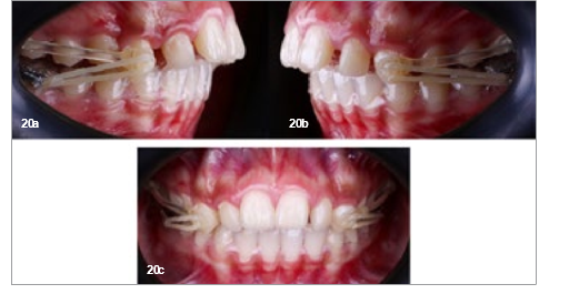
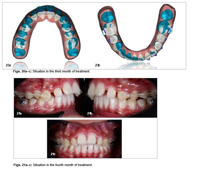
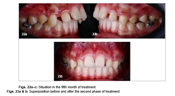
Following the placement of the Carriere Motion Appliances, using our MTO system, both appointments and chair time for the patient are minimised, as no further review is conducted until the third month, when progress is assessed (Fig. 20). If satisfactory progress is observed during this visit, the patient is scheduled for the following month. By the fourth month (Fig. 21), if a bilateral Class I occlusion has been achieved at both the molar and canine levels, we proceed to the second phase of treatment.
The next step involves performing a new intra-oral scan with the Carriere Motion Appliances in place. However, technicians are instructed to virtually remove these. It is crucial to avoid physically removing the Carriere Motion Appliances, as this could compromise the Class I occlusion achieved, leading to relapse. During this phase, the patient continues to wear the Carriere Motion Appliances and the last aligner. Elastics (6.5 oz, ¼ in.) are only worn at night, serving as anchorage until the first aligners for the second phase of treatment arrive (Fig. 22).
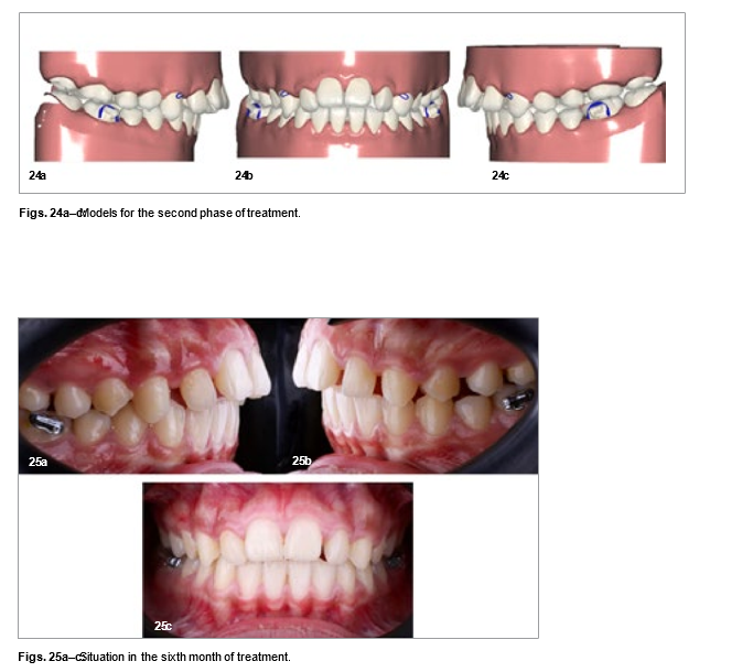
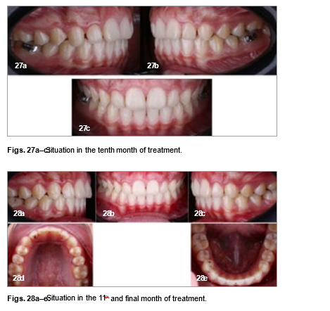
For the second phase of treatment (Figs. 23 & 24), we had the technician design the aligner sequence to close the remaining maxillary spaces through distalisation and retraction of the anterior teeth, applying lingual crown torque to the maxillary incisors and incorporating Class II elastics. To ensure the retention of the Class I occlusion, we instructed the patient to wear Class II elastics (2.5 oz, 3⁄16 in.) nightly. This strategy aimed at closing the maxillary anterior spaces by crown retraction and loss of torque, facilitating relative extrusion to improve the maxillary incisor exposure (Figs. 25–28).
Treatment outcome
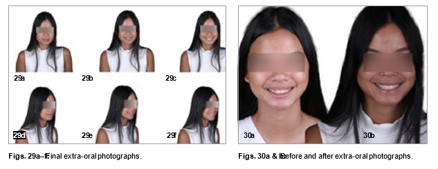
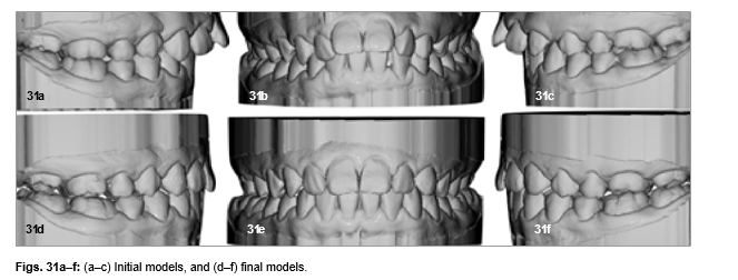
The outcomes proved highly satisfactory for the patient, meeting all the treatment objectives. Noteworthy skeletal alterations were not evident, as the majority of changes observed were primarily dentoalveolar in nature, and the enhancement in the positioning of the maxillary incisors did not affect the patient’s facial profile. From an anterior– posterior perspective, the bilateral Class II malocclusion had been successfully rectified, along with the correction of the overjet (Figs. 29–32). The final transversal arch dimensions attained displayed parabolic and symmetrical arches, demonstrating excellent alignment. The cephalometric measurements displayed minor variations, likely attributable to the patient’s ongoing growth phase, given her stage of cervical maturation (Fig. 33).
The patient presented with a Bolton discrepancy, having excess of the mandibular arch due to the peg-shaped maxillary lateral incisors. Because of the young age of the patient and aesthetic requirements, we decided on bonding veneers on teeth #12 and 22 in a future prosthetic phase instead of mandibular interproximal reduction.
Conclusion
The MTO system stands out as a highly effective and efficient approach for addressing severe Class II cases, providing an alternative to extraction and significantly reducing treatment time, appointments and chair time. In cases of Class II malocclusion that require numerous aligners or aligner stages, the adoption of hybrid treatments such as the one we offer emerges as an advantageous strategy. Within a period of four to five months, we can correct the Class II malocclusion using the Carriere Motion Appliance—this represents the most time-consuming part of the treatment—and then proceed to the Class I phase using aligners to successfully conclude the case.
ABOUT THE AUTHORS:
Dr Carlos Colino holds a master’s degree in dentofacial orthopaedics and clinical orthodontics from the Catholic University of Murcia in Spain and is an associate teacher at the same university. He won first prize for best clinical case at the VIII Congreso Internacional de Alineadores in 2023. He runs his own practice in Malaga in Spain.
Dr Laura Castillo earned her master’s degree in orthodontics from the University of Alcalá in Alcalá de Henares in Spain. She is an associate professor in the orthodontics and dentofacial ortho- paedics master’s programme at the Catholic University of Murcia in Spain. She practises orthodontics exclusively at Dental Colino clinic in Villanueva de la Serena in Spain and is a member of the European Orthodontic Society.
Dr Isabel Drewling undertook her dental degree in 2006 at the University of Barcelona in Spain. She continued her orthodontic and dentofacial orthopaedic training in 2013, and since then, her practice has been limited to orthodontic treatment.
Dr Pedro Colino obtained his PhD summa cum laude from the University of Extremadura in Badajoz in Spain and his MSD in orthodontic and dentofacial orthopaedics with extraordinary award from the University of Alcalá in Alcalá de Henares in Spain. He is head of the orthodontics and dentofacial orthopaedics master’s programme at the Catholic University of Murcia and an associate professor at the Miguel de Cervantes European University in Valladolid, both in Spain. Dr Colino is the author of three orthodontic textbooks and an active member of the Spanish Society of Orthodontics and the European Orthodontic Society. He maintains two private practices in Spain, one in Villanueva de la Serena and another one in Malaga.