
Dr. Ingo Frank
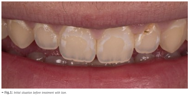
White spot lesions are early signs of demineralization under an apparently intact enamel surface layer. These early enamel lesions show a whitish appearance as a result of an increased porosity within the lesion due to mineral loss [1].
In case of poor oral hygiene or salivary hypofunction even on buccal surfaces of the teeth a plaque accumulation can result in white spot lesions [2]. Especially in patients that underwent orthodontic treatment with brackets white spot lesions can occur due to the difficulties to clean the area adjacent to the bracket. Several clinical studies show a high prevalence of white spot lesions after bracket removal [3, 4].
With preventive strategies like improvement of the oral hygiene and topical fluoride application there is a good chance of arresting early lesions. Though the caries progression may be stopped the whitish appearance often remains as the remineralization is superficial and there is still a porous lesion body underneath [2]. In addition to that stains can be incorporated into the lesion with the result of a brownish appearance of the lesion (brown spots) which often leads to even more esthetic deficiencies. That is why the dentist may become confronted with the patients desire to rehabilitate esthetics. Treating non-cavitated white spot lesions may include tooth bleaching, micro-abrasion, composite fillings or even prosthetic restorations like veneers or combinations of these treatments [5]. All these options are quite invasive implying tooth structure loss. As a micro-invasive alternative caries infiltration (Icon) can be applied to prevent further caries progression. It imbeds the adjunctive effect of masking the whitish appearance of the lesions.
With the infiltrant the porosities in the lesion body are occluded. Therefore, this treatment may be used not only to arrest enamel lesions but also to improve the esthetic appearance of buccal white spots.
Clinical case report
A 19 year-old male patient complained about the appearance of his upper front teeth due to whitish lesions on the vestibular surfaces. He had an orthodontic treatment with brackets during adolescence and when the brackets were removed the white spot lesions became apparent.
In order to fulfill the patient´s desire of an esthetic improvement we suggested an Icon treatment to mask the lesions.
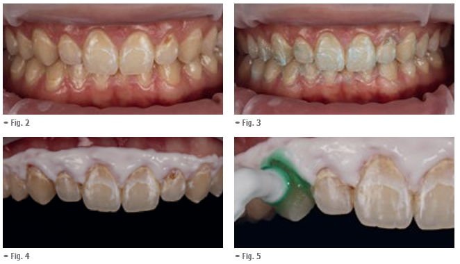
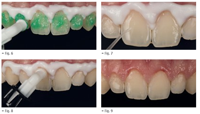
Fig. 2: In order to get an accessible isolated working field optragate was applied.
Fig. 3: Professional cleaning of the vestibular surfaces with fluoride free prophylaxis paste.
Fig. 4: For the application
of Icon an optimal dry working field is mandatory. Furthermore any contact of the material with the gingiva should be avoided. In this case the liquid dam was applied.
Fig. 5: Application of the Icon-Etch for 2 minutes to condition the lesion surface. Fig. 6: The etching gel is covering all white spot lesions of the vestibular surface of the teeth 13-23. The cavitated lesion on the vestibular surface of tooth 22 has been treated with a composite filling. After 2 minutes the etching gel is removed with water spray for 30 seconds and the surface is dried.
Fig. 7: When Icon-Dry is applied, the lesions disappear for the wetting moment. This gives the operator a preview of the results. If the whitish-opaque coloration on the etched enamel diminishes, the procedure can be continued with the infiltrant application. If the lesions are still visible, the etching step should be repeated. After 30 seconds the surfaces are thoroughly dried with oil free and water-free air.
Fig. 8: Application of the infiltrant.
Fig. 9: After the infiltrant has been applied on all treated surfaces, the infiltrant is set for 3 minutes. Excess material is removed with a cotton wad and dental floss before it is light cured for 40s.This infiltrant step is repeated letting the infiltrant set again for 1 minute before excess removal and light curing. Finally the surfaces are polished.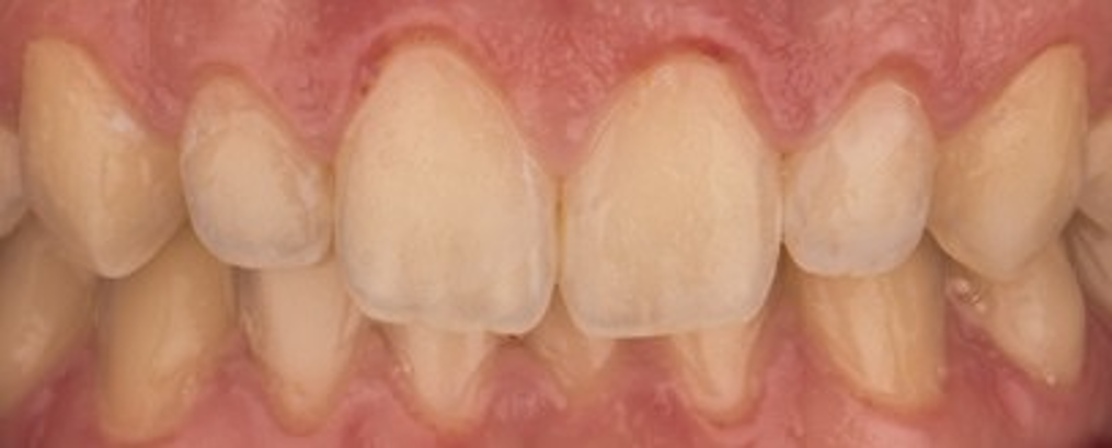
-
- Fig. 10: Final result after Icon treatment. The white spot lesions are masked and a highly satisfying result with large improvement of the esthetic appearance could be achieved.
Discussion
The manifestation of white spot lesions after bracket removal is a common side effect due to the impeded oral hygiene adjacent to the bracket during orthodontic treatment [3, 4, 6]. As esthetic demands of the patients arise dentists do not only have to take care of preventing a further progression of the lesions but also have to deal with the patient´s wishes of masking these lesions which can be esthetic compromising. In contrast to other treatment options like composite filling, micro-abrasion or bleaching, Icon offers a micro-invasive treatment tool without drilling that can not only stop the lesions progressions but also can mask the whitish appearance of the white spot lesions. In case of adjacent cavitated lesions Icon can be successfully combined with a conventional filling in this area. When the cavitated lesion is restricted to enamel the infiltrant even enhances the shear bond strength of the adhesive [7]. Filling procedure and infiltration can be combined in one step. When the cavitation involves dentin the filling should be performed before the Icon treatment as the hydrochloric acid of the Icon-Etch might lead to a decrease of shear bond strength of the adhesive to dentin [8].
Conclusion
With Icon white spot lesions can be masked effectively. It is micro-invasive to the treated enamel surfaces, prevents further demineralization and is easy in handling and application. In case of white spot lesions occurring next to cavitated lesions Icon can successfully be combined with a conventional filling of the cavitated lesion.
Key Learnings
- Infiltration treatment can be combined with direct composite restoration when both white spots and cavitated caries lesion exist on enamel.
- When the cavitated lesion is restricted to enamel the infiltrant even enhances the shear bond strength of the adhesive.
- Filling procedure and infiltration can be combined in one step.
- When the cavitation involves dentin, the filling should be performed before the Icon treatment as the hydrochloric acid of the Icon-Etch might lead to a decrease of shear bond strength of the adhesive to dentin.
-
-
References
- Lee JH, Kim DG, Park CJ, Cho LR. Minimally invasive treatment for esthetic enhancement of white spot lesion in adjacent tooth. The journal of advanced prosthodontics. 2013;5(3):359-63.
- Paris S, Meyer-Lueckel H. Masking of labial enamel white spot lesions by resin infiltration – a clinical report. Quintessence international. 2009;40(9):713-8.
- Gorelick L, Geiger AM, Gwinnett AJ. Incidence of white spot formation after bonding and banding. American journal of orthodontics. 1982;81(2):93-8.
- Hadler-Olsen S, Sandvik K, El-Agroudi MA, Ogaard B. The incidence of caries and white spot lesions in orthodontically treated adolescents with a comprehensive caries prophylactic regimen – a prospective study. European journal of orthodontics. 2012;34(5):633-9.
- Kim S, Kim EY, Jeong TS, Kim JW. The evaluation of resin infiltration for masking labial enamel white spot lesions. International journal of paediatric dentistry / the British Paedodontic Society [and] the International Association of Dentistry for Children. 2011;21(4):241-8.
- Richter AE, Arruda AO, Peters MC, Sohn W. Incidence of caries lesions among patients treated with comprehensive orthodontics. American journal of orthodontics and dentofacial orthopedics : official publication of the American Association of Orthodontists, its constituent societies, and the American Board of Orthodontics. 2011;139(5):657-64.
- Jia L, Stawarczyk B, Schmidlin PR, Attin T, Wiegand A.Effect of caries infiltrant application on shear bond strength of different adhesive systems to sound and demineralized enamel. J Adhes Dent. 2012 Dec;14(6):569-74.
- Jia L, Stawarczyk B, Schmidlin PR, Attin T, Wiegand A. Influence of caries infiltrant contamination on shear bond strength of different adhesives to dentin. Clinical oral investigations. 2013;17(2):643-8.